Peripheral Interventions & Vascular Surgery
Discover the future of vascular imaging with the Kinepict Medical Imaging Tool (KMIT). Specially designed for peripheral interventions and vascular surgeries, KMIT delivers unmatched clarity and precision in imaging small vessels and stenosis. This advanced technology supports healthcare professionals in making accurate, real-time decisions, significantly improving patient outcomes.
KMIT's innovative design reduces the need for iodinated contrast media and minimizes X-ray doses, enhancing patient safety and streamlining workflow efficiency. Whether it's for diagnosing peripheral arterial disease or guiding complex vascular procedures, KMIT is your trusted partner in delivering superior care.
Interventional Oncology & Embolization Procedures
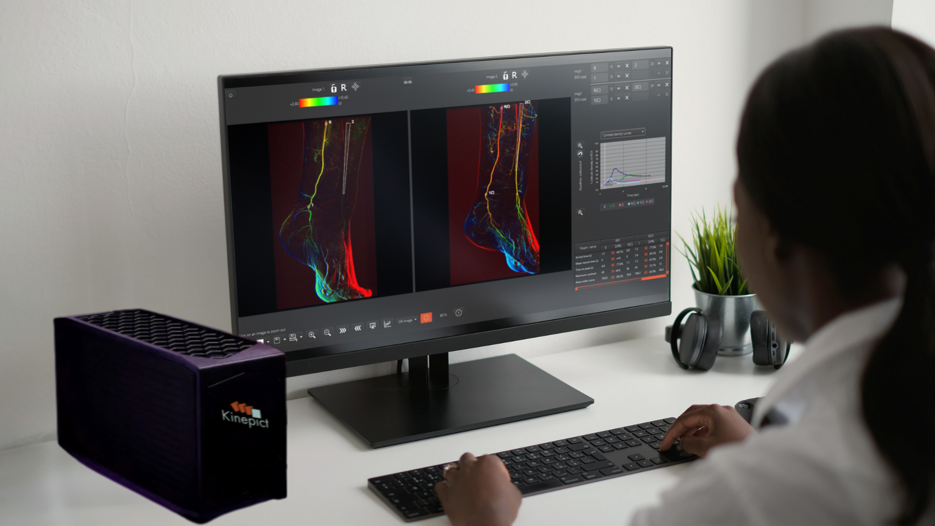
Clinicians trust the Kinepict Medical Imaging Tool (KMIT) for its precise visualization of small vessels in critical procedures such as Uterine Fibroid Embolization and Prostate or Liver Artery Embolization. With advanced features like colorized flow visualization and exceptional image quality, KMIT enables confident decision-making and precise treatment.
KMIT's cutting-edge technology significantly enhances patient safety and cost-effectiveness by reducing the use of Iodinated Contrast Media. This reduction not only minimizes patient risk but also optimizes procedural efficiency.
The benefits.
-
X-ray dose reduction
Studies show that DVA significantly reduces DSA-related radiation exposure during lower limb interventions, thereby enhancing the safety of endovascular procedures. This technology offers considerable radiation reduction benefits for both patients and medical staff. The clinical findings have been published in esteemed peer-reviewed journals.
-
ICM dose reduction
Clinical studies reveal that the technology's quality reserve can be leveraged to substantially reduce contrast media during angiography procedures. This reduction offers significant economic and health benefits, including fewer adverse events and a decreased risk of contrast-induced nephropathy for patients, while also providing cost savings for cath labs. The study was published in the European Journal of Radiology Open.
-
Increased diagnostic insight
DVA provides enhanced visualization of the same structures as DSA, offering superior image quality (Gyano et al., 2019; Bastian et al., 2021; Thomas et al., 2021). Noteworthy is DVA's ability to improve the image quality of angiograms in CO2 angiography (Orias et al., 2019). This enhancement is particularly essential as CO2 serves as the sole viable contrast agent for patients with severe kidney injury. Even at lower frame rates (1–3 fps), CO2 angiography benefits from improved image quality, leading to significant reductions in radiation exposure (Gyano et al., 2020).
See the difference.
How it works.
Transform x-ray technology through cutting-edge data analytics with only 3 steps.
Lorem ipsum dolor amet prism banjo beard brooklyn vaporware vexillologist. Swag cardigan PBR&B affogato, health goth vice.
Sanda Cambel
Director of Marketing
Lorem ipsum dolor amet prism banjo beard brooklyn vaporware vexillologist. Swag cardigan PBR&B affogato, health goth vice.
Sanda Cambel
Director of Marketing
Lorem ipsum dolor amet prism banjo beard brooklyn vaporware vexillologist. Swag cardigan PBR&B affogato, health goth vice.